Welcome to the Amira-Avizo Software Use Case Gallery
Below you will find a collection of use cases of our 3D data visualization and analysis software. These use cases include scientific publications, articles, papers, posters, presentations or even videos that show how Amira-Avizo Software is used to address various scientific and industrial research topics.
Use the Domain selector to filter by main application area, and use the Search box to enter keywords related to specific topics you are interested in.
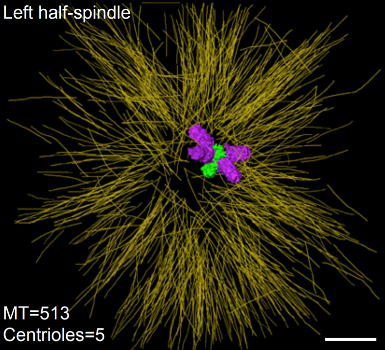
Asymmetric Centriole Numbers at Spindle Poles Cause Chromosome Missegregation in Cancer
Chromosomal instability is a hallmark of cancer and correlates with the presence of extra centrosomes, which originate from centriole overduplication.
Overduplicated centrioles lead to the formation of centriole rosettes, which mature into supernumerary centrosomes in the subsequent cell cycle. While extra centrosomes promote chromosome missegregation by clustering into pseudo-bipolar spindles, the contribution of centriole rosettes to chromosome missegregation is unknown. We us... Read more
Marco R.Cosenza, Anna Cazzola, Annik Rossberg, Nicole L. Schieber, Gleb Konotop, Elena Bausch, Alla Slynko, Tim Holland-Letz, Marc S.Raab, Taronish Dubash, Hanno Glimm, Sven Poppelreuther, Christel Herold-Mende, Yannick Schwab, Alwin Krämer
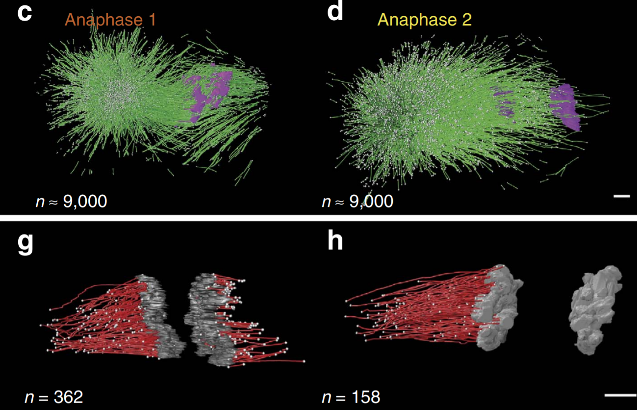
C. elegans chromosomes connect to centrosomes by anchoring into the spindle network
The mitotic spindle ensures the faithful segregation of chromosomes. Here we combine the first large-scale serial electron tomography of whole mitotic spindles in early C. elegans embryos with live-cell imaging to reconstruct all microtubules in 3D and identify their plus- and minus-ends. We classify them as kinetochore (KMTs), spindle (SMTs) or astral microtubules (AMTs) according to their positions, and quantify distinct properties of each class. While our light microscopy and muta... Read more
Stefanie Redemann, Johannes Baumgart, Norbert Lindow, Michael Shelley, Ehssan Nazockdast, Andrea Kratz, Steffen Prohaska, Jan Brugués, Sebastian Fürthauer & Thomas Müller-Reichert
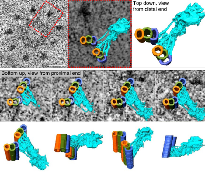
Centrioles are vital cellular structures that form centrosomes and cilia. The formation and function of cilia depends on a set of centriole’s distal appendages. In this study, we use correlative super resolution and electron microscopy to precisely determine where distal appendage proteins localize in relation to the centriole microtubules and appendage electron densities. Here we characterize a novel distal appendage protein ANKRD26 and detail, in high resolution, the initial steps of dist... Read more
Mathew Bowler, Dong Kong, Shufeng Sun, Rashmi Nanjundappa, Lauren Evans, Veronica Farmer, Andrew Holland, Moe R. Mahjoub, Haixin Sui & Jadranka Loncarek

Soluble tubulin is locally enriched at mitotic centrosomes in C. elegans
During mitosis, the centrosome expands its capacity to nucleate microtubules. Understanding the mechanisms of centrosomal microtubule nucleation is, however, constrained by a lack of knowledge of the amount of soluble and polymer tubulin at mitotic centrosomes. Here we combined light microscopy and serial-section electron tomography to measure the amount of dimer and polymer at mitotic centrosomes in early C. elegans embryos. We show that a C. elegans one-cell stage centrosome at metaphase co... Read more
Johannes Baumgart, Marcel Kirchner, Stefanie Redemann, Jeffrey Woodruff, Jean-Marc Verbavatz, Frank Julicher, Anthony Hyman, Thomas Mueller-Reichert, Jan Brugues
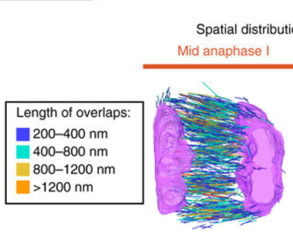
Chromosome segregation occurs by microtubule pushing in oocytes
During cell division, spindle microtubules ensure an equal repartition of chromosomes between the two daughter cells. While the kinetochore-dependent mechanisms that drive mitotic chromosome segregation are well understood, in oocytes of most species atypical spindles assembled in absence of centrosomes entail poorly understood mechanisms of chromosome segregation. In particular, the structure(s) responsible for force generation during meiotic chromosome separation in oocytes is unclear. Usin... Read more
Kimberley Laband, Rémi Le Borgne, Frances Edwards, Marine Stefanutti, Julie C. Canman, Jean-Marc Verbavatz, Julien Dumont